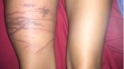
Philadelphia, September 5, 2019 - A detailed case report and comprehensive sequence of photographs in Wilderness & Environmental Medicine, published by Elsevier, document the dermatological progression of a patient stung by a jellyfish off the coast of Cambodia. The aim of this report is to guide clinicians and patients to understand what to expect after such a sting and steps to increase the probability of a full recovery.
Jellyfish stings (envenomations) are a common affliction of ocean goers worldwide. Although many are simply a nuisance, some can be very severe or even fatal. The most severe non-anaphylactic reactions are caused by jellyfish species that inhabit Indo-Pacific waters. Examples of species known to be clinically dangerous include the box jellyfish (Chironex fleckeri), sea nettle (Chrysaora quinquecirrha), and Irukandji (Carukia barnesi).
"In this report, we provide written and visual details of the natural progression of a probable box jellyfish envenomation that originated in waters off Cambodia," explained lead author Paul S. Auerbach, MD, Department of Emergency Medicine, Stanford University School of Medicine, Palo Alto, CA, USA. "We document a comprehensive visual demonstration of what patients and clinicians should expect after a severe sting by box jellyfish or similarly injurious species."
The patient (a co-author of this report) was a 21-year-old woman in good health at the time of injury in early June 2019. She was stung while swimming off the coast of Koh Ta Kiev, Cambodia, in chest-deep water. She experienced immediate and intense burning pain to her right leg that gradually subsided over the next 10 hours, bright red and swollen streaks where tentacles had contacted the skin of the right thigh, and lightheadedness with a brief loss of consciousness after leaving the water within 20 minutes of the sting. She did not see the jellyfish well enough to identify precisely the species, but multiple species of box-type jellyfish are known to live in these waters.
With worsening symptoms, the patient travelled to Siem Reap where she was admitted to Royal Angkor International Hospital overnight. She then travelled to Bali. Dr. Auerbach telephoned the patient and recommended that she return to California promptly to receive the medical care necessary to attempt healing without complications and gave her detailed advice for managing the wound during the journey.
After arriving in the United States, the patient was seen by a dive medicine specialist physician and a plastic surgeon. Physical examination was consistent with superficial and deep partial thickness burns, possibly with near full thickness injury in some areas.
She was advised to continue washing the wounds and to clean and dress the wounds using silver sulfadiazine topical cream in the deeper areas and Leptospermum honey (Medihoney) in the shallower areas. The immediate goals were to keep the wounds clean, prevent infection, maintain moisture to facilitate healing, and allow for serial examinations to determine the best method for wound closure.
After 60 days the wound was fully closed and healed. Moisturizing lotion and sun protection measures were continued to optimize scar maturation. The patient was counseled that she might be a candidate for elective scar revision, steroid treatment, and/or laser therapy in the future.
"With knowledgeable consultation from experienced physicians and meticulous care, this injury healed without the need for skin grafting," reported Dr. Auerbach.
"The authors have provided an exceptionally detailed photographic timeline of the case," added Neal W. Pollock, PhD, Department of Kinesiology, Laval University, Quebec, QC, Canada, and Editor-in-Chief of Wilderness & Environmental Medicine. "It provides excellent documentation of the evolution that will rarely be seen."


